櫟山螢雪研究所
When that I was and a little tiny boy,
   With hey, ho, the wind and the rain,
A foolish thing was but a toy,
   For the rain it raineth every day.
  ••••••••••••••••••••••••••••••••••
A great while ago the world begun,
   With hey, ho, the wind and the rain,
But that's all one, our play is done,
   And we'll strive to please you every day.

                               from Twelfth Night


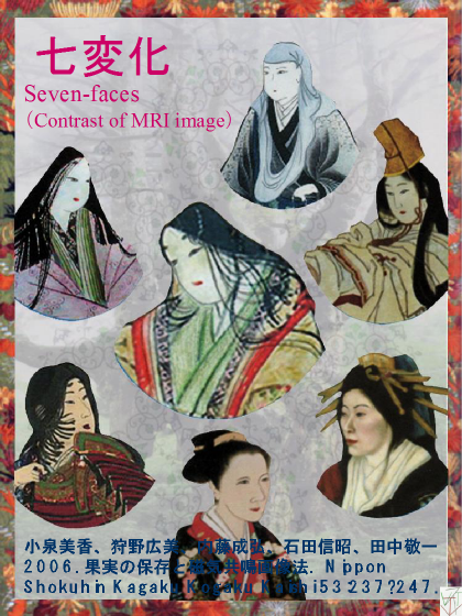
Seven-faces


 物を観測するとき、一義的に対象の形が決まり何であるかを同定することができると期待しがちである。しかし、それはMRIの測定には当てはまらない。MRIは、生物体内の構造を非破壊的に観察する(Hills, 1998; McCarthy, 1994; Wanget al., 1988; WangandWang, 1989)ために開発された。しかし、実際には、体内形態だけでなく、同一対象のイメージが測定法および測定条件によって著しく変化することを基に、組織の生理や性質について多くの情報を得られるところに意味がある。
 MRIを始めたころ、測定条件を変えると著しく異なったイメージが取れることに面食らい、慣れるのには長い時間を費やさなければならなかった。MRIにおいて、得られる一次情報はイメージにおけるシグナルの強弱である、コントラストと言われている。コントラストは測定対象の性質だけでなく、測定条件と内部環境によって著しい変化を示し、その本質的意味が解らないとイメージから充分な情報を得ることが出来ない。しかし、初心者の時代を振り返ると、理論を理解するだけで疲れてしまってMRIは実用になるのか?と疑問を持ったことを覚えている。

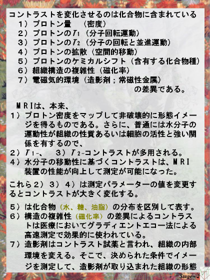
 コントラストを変化させるのは、化合物の
  1) プロトン量 (密度)
  2) プロトンのT1 (分子回転運動)
  3) プロトンのT2 (分子の回転と並進運動)
  4) プロトンの空間的移動 (拡散とフロー)
  5) プロトンを含有する化合物種 (ケミカルシフト)
  6) 組織構造の複雑性 (磁化率)
  7) 電磁気的環境(造影剤;常磁性金属)
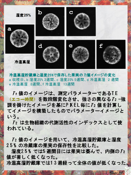
の差異である。イメージングは、本来、1)プロトン密度をマップして非破壊的に形態イメージを得るものである。さらに、普通には水分子の運動性が組織の性質あるいは細胞の活性と強い関係を有するので、2)T 1-、3)T 2-コントラストが多用される。4)水分子の移動性に基づくコントラストは、 MRI装置の性能が向上して測定が可能になった。これら2)3)4)は測定パラメーターの値を変更するとコントラストが大きく変化する性質を持つ(Ishidaet al., 2000)。ここでは、ブルーベリーを試料として、これらの測定法が果実の収穫後生理の研究手法として用いられている意味を説明した(Clarket al., 1997; Hills, 1998; Koizumiet al., 2000)。

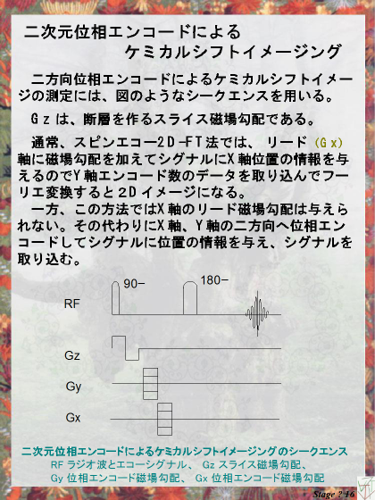
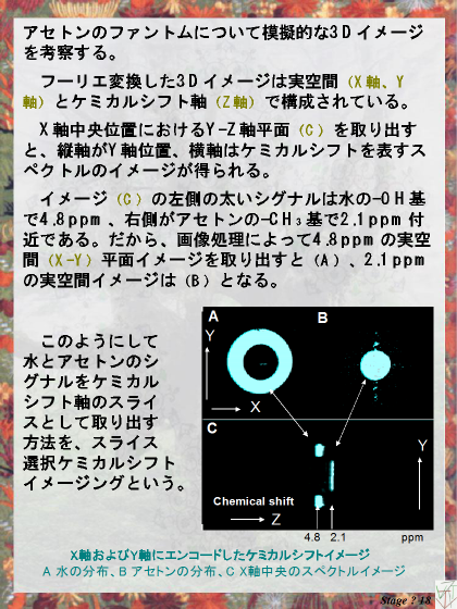
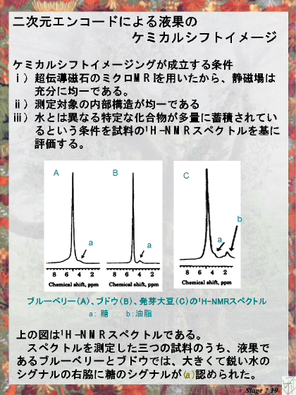
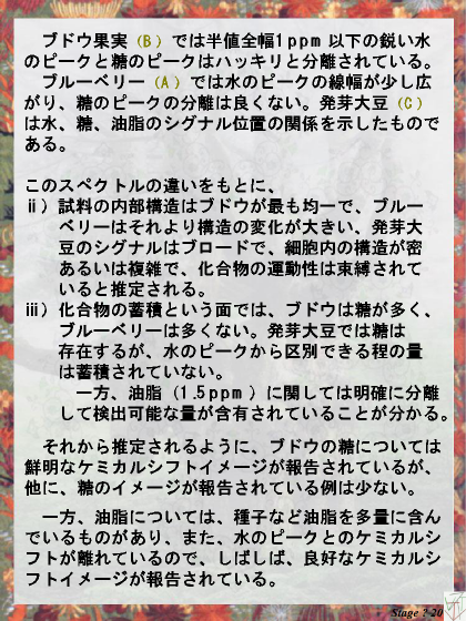
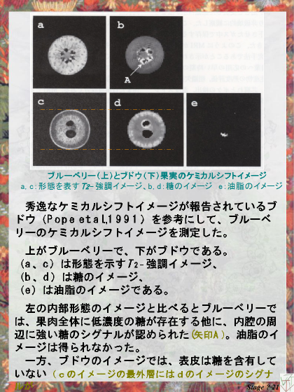
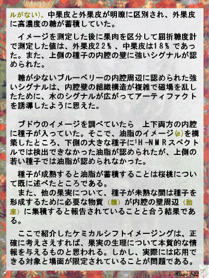
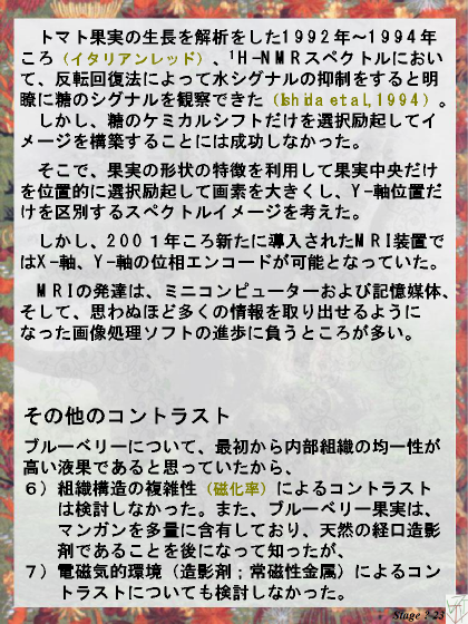
 5)ケミカルシフトイメージングは異なる化合物(水、糖、油脂)の分布を区別して表す方法である。高い技術を必要とするので、1990年初頭には測定法の工夫に魅力を感じた(Bottomleyet al., 1986; Ishidaet al., 2000)が、2000年頃に導入された新しい装置では、メーカーによって二次元位相エンコードによる測定シークエンスプログラムが用意されていた(Brownet al., 1982)。1995年以降のミニコンピューターの発達と性能向上によるものである。ケミカルシフトイメージングは油脂を多量に含有する種子の研究(Gussoniet al., 1993; Halloinet al., 1993; Jagannathanet al., 1995)や、糖が品質の要素となる果実の研究(Pope 1992; RumpelandPope, 1992)では重要な測定法であるから、ブドウおよびブルーベリーを用いて測定法とイメージの意味を説明した。
 七変化では採り上げなかったコントラストが、グラディエントエコー法による高速測定において効果的に使われている6)構造の複雑性(磁化率)の差異によるコントラスト、および、水分子周辺の電磁気学的内部環境を変える7)造影剤によるコントラストである。前者6)は組織構造の性質の違いによるイメージであり、短時間の測定で骨などの形態を明瞭に表す、一方、後者7)造影剤は疾病がある組織に集積して組織の特定ができるというように一意的に意味が定まるので、MRIとして使いやすい方法であるし、決められた条件でイメージを測定して組織障害や造影剤が取り込まれた組織の形態および状態についての情報を得る医療分野の効果的な技術となっている。
 同一の対象を測定したMRIのイメージは、外形が似ていてもイメージが示す意味が異なっていたり、反対に、同一の対象を測定したのに輪郭さえ検出できない場合がある。よって、イメージの解釈には測定法とコントラストの関係を正確に理解することが大切である。
                                                                                  (研究員)
 

文献

果実とMRI
Clark CJ, Hockings PD, Joyce DC, Mazucco RA. 1997. Application of magnetic resonance imaging to pre- and post-harveststudies of fruits and vegetables. Postharv. Biol Technol. 11: 1-21.
 Hills B. 1998. Magnetic resonance imaging in food science. New York, John Wiley & Sons, Inc.
 Koizumi M, Ishida N, Kano H. 2000. Postharvest fruits and MRI. Current Topics Plant Biol 2: 1-20.
 McCarthy MJ. 1994. Magnetic resonance imaging in foods. Chapman & Hall.
 Wang SY, Wang PC, Faust M. 1988. Non-destructive detection of watercore in apple with nuclear magnetic resonanceimaging.Sci Hort 35: 227-234.
 Wang CY, Wang PC. 1989. Nondestructive detection of core breakdown inBartlettpears with nuclear magneticresonanceimaging. HortSci 24: 106-109.

コントラスト
 Callaghan PT. 1991. Principles of nuclear magnetic resonance microscopy. Oxford, Clarendon Press.

ケミカルシフトイメージング
Bottomley PA, Foster TH, Leue WM. 1984.In vivonuclear magnetic resonance chemical shift imaging by selective irradiation.Proc Natl Acad Sci USA. 81:6856-6860.
 Brown TR, Kincaid BM, Ugurbil K. 1982. NMR chemical shift imaging in three dimensions. Proc Natl Acad Sci USA. 79: 3523-3526.
 Gussoni M, Greco F, Consonni R, Molinari H, Zannoni G, Bianchi G, Zetta L. 1993. Application of NMR microscopy tothehistochemistry study of olives (Olea europeaL.). Magn Reson Imaging 11: 259-268.
 McCarthy MJ. 1994. Magnetic resonance imaging in foods. Chapman & Hall.
 Halloin JM, Cooper TG, Potchen EJ, Tompson TE. 1993. Proton magnetic resonance imaging of lipid in pea embryos. J AmOilChem Sci 70: 1259-1262.
 Ishida N, Koizumi M, Kano H. 2000. The NMR microscope: a unique and promising tool for plant science. Annal Bot 86: 259-278.
 Jagannathan NR, Govindaraju V, Raghunathan P. 1995.In vivomagnetic resonance study of the histochemistry of coconut(Cocos nucifera). Magn Reson Imaging 13: 885-892.
 Pope JM. 1992. Applications of chemical shift microscopy to noninvasive histochemistry of plant materials. In BlÜmich B,KuhnW. eds. Magnetic resonance microscopy: methods and applications in materials science, agriculture and biomedicine. NewYork,VCH Publishers, 441-457.
Rumpel H. Pope JM. 1992. The application of 3D chemical shift microscopy to noninvasive histochemistry. Magn ResonImaging10:187-194.

冷温高湿貯蔵庫
 田中敬一,朝倉利員,谷村泰宏.1998. 冷温高湿貯蔵の開拓,冷熱輻射方式貯蔵庫,負イオン/オゾン混合ガスによる果実の鮮度保持.冷凍 73: 58-63.


          A great while ago the world began,
               With hey-ho, the wind and the rain;
           But that's all one, our play is done,
               And we'll strive to please you every day.